CYTO 2014 Notes: Fluorescence Lifetime Workshop
This is the first blog in a series covering CYTO 2014, held this May 17-22 in Ft. Lauderdale, Florida. Mainly in the form of raw notes, the intent of this series is to highlight talks, events, and trends from the annual cytometry congress and trade show that I found noteworthy. Clarifications, corrections, comments, and other suggestions welcome.

The Fluorescence Lifetime Workshop took place Monday, May 19. Co-chaired by Profs. Jessica Houston (New Mexico State University, Las Cruces, NM) and Silas Leavesley (University of South Alabama, Mobile, AL), the workshop featured the following panel of presenters: Patrick Jenkins (New Mexico State University, Las Cruces, NM); Prof. Miho Suzuki (Saitama University, Japan); Prof. János Szöllősi (Debrecen University, Debrecen, Hungary); and Dr. Ralph Jimenez (JILA/NIST and University of Colorado, Boulder, CO).
About 50 people attended the workshop. My notes of the presenters’ talks follow–see also other pictures of the event.
Patrick Jenkins, NMSU
Fluorescence Lifetime Flow CyTAUmetry
– frequency domain FLT FC
– FLT CVs in beads: ~ 5-20%
– FLT CVs in cells: 15-30%
– FLT CVs in yeast: 7%
– yeast: tealFP strain and darkCitrine-tealFP FRET strain
– FLT only difference (2 vs. 3 ns)
heavy spectral overlap and similar FL intensity
– sorted @ 60-99% purity on either strain
Miho Suzuki, Saitama U.
Signal Transduction with FLT FC
– chimeric FRET / FLIM based bioprobes
– based on combining different biomaterials
– organic dyes, FPs, QDs in different pairwise FRET combinations
– cysteine – rare aminoacid
– GFP has two instances of cysteine
one inside the barrel and one on the outside
latter suited for chemical mods
– ID’d two aminoacids in GFP suitable for FRET
– used Flicyme 300 FLT FC
János Szöllősi, Debrecen U.
FLIM & TIRF
– collaborator: György Vereb (fmr student)
– what kind of bio questions need FRET-FLIM?
– ErbB receptor tyrosine kinases and their associations
– problems
– uneven labeling of tissue
– low intensity due to poor labeling locally
– limited usefulness
– ErbB1-4, HER1-4
– ErbB1 also = epidermal growth factor receptor
– ErbB2 – no ligand; if overexpressed, high chance for metastasis
– ErbB3 – no kinase activity
– involved in MAPK and PI3K signaling pathways
– looking at heterogeneity of FLT signal within the cell (pixel by pixel)
– FRET tells you if the proteins have associated
– use Lambert LI-FLIM added to microscope
– Olympus Microscope IX81 and IX2
– Olympus 4-ch. CellTIRF
– motorized stages
– donor only: 3.44 ns; donor acceptor: 3.19 ns;
donor acceptor + EGF: 2.89 ns
– FLT shift under archazolid treatment: ~300 ps (FLT distrib ~ 1 ns wide)
– phasor plot to look at stoichiometry of associated proteins
Ralph Jimenez, JILA/NIST/U. Colorado
Lifetime and Photobleaching for new FPs
– organic dyes ~100x more stable than FPs
– brightness of different FPs peaks at 532 and goes to 20% at 425 and 650
– want to develop brighter, more stable FPs
– photostability increased by O2 reduction
– pulsed photobleaching assays on single HeLa cells (2 ms on, 8 ms off)
– photobleaching needs to be studied at relevant intensities
(10^2-10^5 W/cm2)
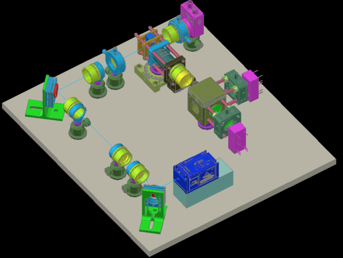
– microfluidic channel with multiple bleaching
532 nm beams in line source config
– use optical trapping with 1064 nm beam to steer cells to “keep” path
– studied mCherry (most photostable in mFruit family)
– found mCherry mutant with 3x higher stability but 2x lower quantum yield
– use additional laser beam to measure lifetime and stability simultaneously
– Q (JS): saturation?
– A (RJ): no, 1 photon every 20 ns
Image credits: CYTO/ISAC