Step-by-Step Protocol for the Delaware
Thanks to our collaboration with Dr. Christopher Ward at the University of Kansas, we’ve published a complete, step-by-step protocol for the analysis of urinary EVs — and with it, we demonstrate how you can achieve *clear, reproducible results* using the Delaware Flow NanoCytometer.
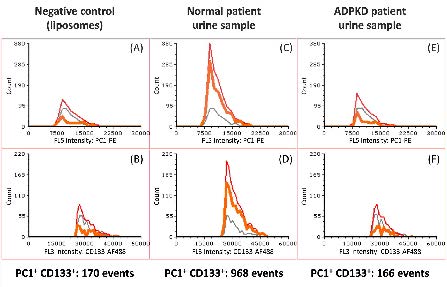
In healthy individuals, urinary vesicles naturally carry the PC1 protein, a key marker inversely correlated with kidney disease.
In patients with autosomal dominant polycystic kidney disease (ADPKD), these PC1-positive vesicles are significantly reduced, and our technology, coupled with Dr. Ward’s antibody markers, reveals this contrast clearly.
The Delaware system allows for:
– fast sample prep (*no* ultracentrifugation)
– multicolor detection (up to 6 colors)
– high sensitivity
– quantitative analysis of PC1 and other EV proteins
This protocol enables researchers to:
– differentiate between healthy and abnormal kidney samples
– apply similar workflows to other minimally processed media (blood, plasma, cell culture, etc.)
– detect EVs with high specificity using multiple protein markers to reduce background
Explore the full protocol in our new Application Note, click here to learn more.
Reach out if you are interested in a demo; our team can run your samples on the Delaware Flow NanoCytometer or help you bring this technology to your lab.