We’re back in Taipei for another install—and this beautiful city is starting to feel like home.
Cytometry Innovations Continue to Surprise Insiders
CYTO 2013 (XXVIII Congress of the International Society for Advancement of Cytometry), the annual “watering-hole” gathering for cytometrists of all shapes and sizes, took place in San Diego this year, and it was exciting as ever. The scientific program turned up a number of interesting new research results, and the trade show floor had the customary line-up of old favorites, plus several intriguing new entrants.
A popular session was “Trends in Cytometry Instrumentation,” a workshop I was honored to co-chair together with Prof. Steven Graves, Associate Director of the Center for Biomedical Engineering at the University of New Mexico in Albuquerque, NM. The topics selected for presentation met the following criteria: (i) the papers exemplified broad technology currents that the workshop co-chairs had identified as important to cytometry, both flow- and image-based; and (ii) the speakers did not already have an oral presentation in the program.
To analyze the instrumentation trends, we examined the “demand side” (application drivers) and the “supply side” (technology enablers), and we came up with the following lists:
- A. Cytometry instrumentation drivers (applications)
– smaller systems
– smaller particles
– greater multiplexing
– richer information - B. Cytometry instrumentation enablers (technology)
– microfluidics
– on-chip sorting
– acoustic cytometry
– higher-power lasers
– dense source wavelength coverage
– higher sensitivity
– spectral resolution
– tunability
– novel probes
– waveform analysis
– fluorescence lifetime
– light scattering
Several research papers were then selected to illustrate many of the enabling technologies and stimulate thoughts and discussions. The result was this outstanding lineup of speakers and technology research:
- Tony Jun Huang, of Pennsylvania State University spoke about sorting by Standing Surface Acoustic Waves in a microfluidic flow cytometer;
- Lu Gao, of Duke University, described on-chip sorting using acoustically enhanced magnetophoresis in a microfluidic cytometer;
- Ola Jakobsson, of Lund University, reported on a new application of acoustic cytometry in orienting red blood cells in flow;
- Peter Favreau, of the University of South Alabama, showed the use of tunable spectral filters in hyperspectral microscopy;
- Martin Büscher, of Miltenyi Biotec, talked about the use of nano-gold Surface Plasmon Resonance particles for more effective multiplexing;
- Mark Naivar, of DarklingX, highlighted the capture of whole waveforms instead of listmode data in flow cytometry and their use in analysis;
- Ruofan Cao, of New Mexico State University (group webpage), discussed integration of fluorescence lifetime detection into standard flow cytometers.
Additionally, the following two papers were selected but could not be presented at the workshop due to schedule conflicts:
- Vasilis Toxavidis, of Beth Israel Deaconess Medical Center (group webpage), with a paper on a modified commercial sorter to detect and harvest microparticles;
- Anastasiya Konokhova, of Novosibirsk State University (group webpage), with research describing Scanning Flow Cytometry to characterize E. coli.
Each of the speakers was asked, in presenting the material, to address three key points:
- What are the advantages of your approach for the intended application?
- What is the greatest barrier to widespread adoption of this technology?
- How will this innovation translate to the clinical or research environment?
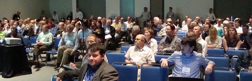
Fig. 1. Attendees to the Trends in Cytometry Instrumentation workshop,
with co-chair Prof. Steve Graves in the foreground.
The session was very well attended (see Fig. 1), followed by a lively discussion. The take-away was that flow and image cytometry, while benefiting from decades of refinement and technology maturation, are continually surprising us with new ways to solve existing and emerging problems.
For example, several of the speakers discussed chip-based microflow technologies in conjunction with acoustic forces. The growing number of groups working in this area is making the possibility of sheathless, much more compact flow cytometers increasingly real. Likewise there has been an explosion in research and development aimed at detecting microparticles—increasingly recognized as important diagnostic biomarkers, whereas just a few years ago even their existence was not widely known. And the showcased innovations on the signal side (through novel probes and scattering analysis, hyperspectral techniques, fluorescence lifetime, and richer full-waveform data sets) indicate that the final word on multiplexing is far from having been written.
Looking forward to next year’s edition!
Image credit: G. Vacca
Related Posts

Comments (0)