DELAWARE
Detection and characterization of sub-micron entities, including extracellular vesicles (EVs) and exosomes, represents an important next frontier in both research and clinical applications. These nanoparticles produce exceedingly small scattering and fluorescent signals which standard commercial flow cytometers cannot detect. Even systems designed to address this application have, thus far, fallen short, creating an unmet and growing demand for a nanoparticle analysis system with suitable usability and throughput.
We designed and developed the Delaware Flow NanoCytometer® specifically to meet the demanding needs of nanoparticle researchers, providing sensitive detection and characterization of biological and non- biological nanoparticles. Based on our modular, customizable Potomac architecture, the system incorporates design modifications intended to enhance nanoparticle sensitivity without compromising throughput.

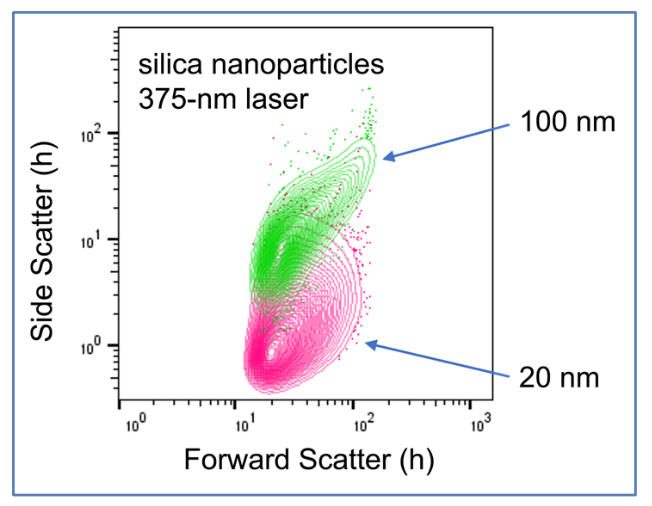
Silica nanoparticles run on the Delaware
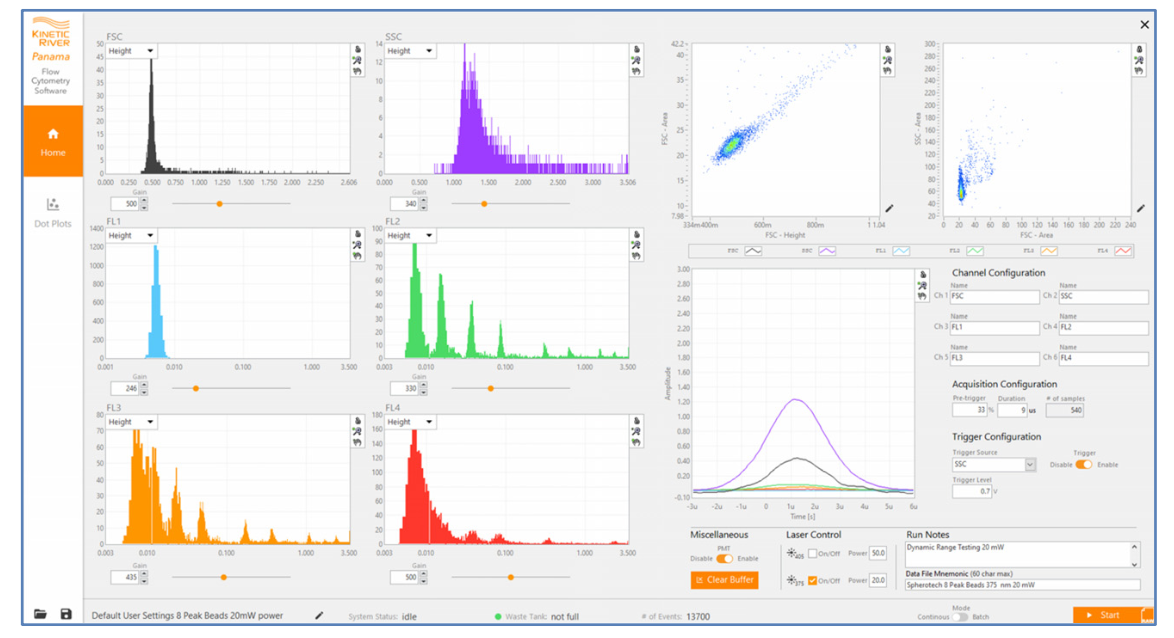
The Panama software for instrument control and data visualization
The Delaware’s high-power lasers provide up to five excitation wavelengths (375, 405, 488, 561, and 640 nm) and a proprietary high-NA collection lens delivers maximum sensitivity. The system offers up to three scattering channels and up to six fluorescence detection channels. The Delaware features Kinetic River’s Shasta fluidic control system for ultrastable sheath flow and superior core stream control. The Cavour always-on flowcell monitor allows you to optimize laser alignment and core stream dimensions in real-time without removing the cover. The entire system is operated using our intuitive, easy-to-use Panama flow cytometry software for instrument control and data visualization, providing researchers with the flexibility their cutting-edge research requires.
This carefully-crafted instrument has been extensively tested on a variety of materials including polystyrene (down to 60 nm) and colloidal silica (down to 100 nm) nanoparticles, fluorescent nanospheres (100 nm), hollow organo silica beads (374 nm) and lipoprotein shells (100 nm), demonstrating sensitivity to at least 60 nm to meet some of the most demanding applications. The Delaware Flow NanoCytometer combines ease of use with advanced nanoparticle sensitivity to offer users a powerful new tool for exosome and EV research.
The Delaware – see what you’ve been missing.

Best-in-class resolution. Kits of highly monodisperse polystyrene nanoparticles (Rosetta Beads, Exometry) ranging from 100 nm to 400 nm were analyzed on the Delaware. The resulting clusters (left panel) are very tight, easily allowing identification of the 100-, 125-, and 147-nm subpopulations (the 150-nm cluster intersperses due to its fluorescence, which alters the effective refractive index). Analyzing the scattering spread of individual populations (right panel), we determine a minimum spread of 6 nm in the 125-nm cluster—an unprecedented result.

Multicolor Analysis. We analyzed HEK293 cell-derived exosomes (FLuo-EVs, HansaBioMed) engineered to express EGFP. By NTA (ZetaView, Particle Metrix), these EVs have an average size of 90 nm. On the Delaware, they show up clearly by scattering (left panel), by the endogenously expressed fluorescence (middle panel), and by cell membrane staining (right panel). We have additionally stained them with fluors conjugated to CD9, CD63, CD81.

Clog-free operation from 28 nm to 28 μm. The Delaware can handle very heterogeneous samples, with particle sizes spanning more than one order of magnitude. It does not clog when particles larger than those in the target nanometer range pass through the flowcell. It can even switch from analyzing very small nanoparticles (left panel) to running conventional cell assays on PBMCs or larger cells (middle and right panels).
Performance
Nanoparticle detection (375-, 405-, and 488-nm excitation; 3 scattering channels):
- 60-nm Spherotech polystyrene
- better than 100 nm Alpha Nanotech colloidal silica
EV surrogates:
- 100-nm Cellarcus lipoprotein shells
- 374-nm Exometry Verity shells
Dynamic range (375-, 405-, 488-nm excitation):
- high sensitivity: approx. 60 nm to 300 nm (PS)
- approx. 100 nm to 1µm (silica)
Specifications
Configurations
Basic Configuration
(Tier 3)
2 Lasers
405 nm, 300 mW*
488 nm, 300 mW*
*pre-fiber
Standard Scattering
FSC, SSC
(405 and 488 nm)
2 Fluorescence Channels*
531/42
573/14
*All filters user-removable and -replaceable
High Sensitivity Configuration
(Tier 2)
3 Lasers
375 nm, 70 mW
405 nm, 300 mW*
488 nm, 300 mW*
*pre-fiber
Ultrasensitive Scattering
FSC, SSC
(375, 405, 488 nm)
4 Fluorescence Channels*
531/42
573/14
624/28
697/42
*All filters user-removable and -replaceable
Five-Laser Configuration
(Tier 1)
5 Lasers
375 nm, 70 mW
405 nm, 300 mW*
488 nm, 300 mW*
561 nm, 200 mW
640 nm, 100 mW
*pre-fiber
Ultrasensitive Scattering
FSC, SSC
(375, 405, 488, 561, 640 nm)
6 Fluorescence Channels*
440/40†
531/42
573/14
624/28
697/42
755 LP
*All filters user-removable and -replaceable
† Alternate to SSC 2
| Tier 3 (Basic Configuration) Specifications | ||
|---|---|---|
| Laser 1 | Wavelength | 405 nm |
| Power | 300 mW pre-fiber | |
| Laser 2 | Wavelength | 488 nm |
| Power | 300 mW pre-fiber | |
| Detector 1 | Forward-scatter | 488 nm |
| Detector 2 | Side-scatter 1 | 405 and 488 nm |
| Detector 3 | Fluorescence 2 | 510–552 (531/42) nm |
| Detector 4 | Fluorescence 3 | 566–580 (573/14) nm |
| Shasta Pneumatics & Fluidics Control Module | Sheath pressure (primary) | 10 psi (factory set) |
| Sample pressure (primary) | 10 psi (factory set) | |
| Sheath pressure (secondary) | 2–8 psi, user-settable | |
| Sample pressure (secondary) | 2–4 psi, user-settable | |
| Variable sample injection flow rate | 5–100 μL/min | |
| Ultrafiltered sheath fluid container | 9.5 L | |
| Waste container | 9.5 L | |
| Cavour Flowcell Monitoring Module | Always-on display | Dedicated second monitor |
| Field of view | Includes flowcell channel and inner sidewalls at point of laser intercepts | |
| Focusing | User adjustable | |
| Reduction of scattered light | 520 nm longpass (user-removable/replaceable) | |
| Data logging | Photo, video, calibrated on-screen measurements | |
| Panama Flow Cytometry Software | User laser control (on/off, power levels for each laser) | |
| User PMT control (global on/off, real-time gain setting for each channel) | ||
| User-settable event trigger parameters | ||
| Live oscilloscope traces for all active channels | ||
| Live, customizable histograms for all active channels with option to display background curves | ||
| Live, customizable dot plots for all bivariate channel combinations with option to display background populations | ||
| Live, automatic, Mie-theory particle size distribution histogram for selected particle types | ||
| Real-time sample flow rate, absolute event count, absolute concentration indicators | ||
| Waste full alert | ||
| FCS 3.1 data format export (listmode format with timestamp, height, area, width for each event on all active channels) | ||
| Physical | Weight (excluding monitors, monitor stands, keyboard, mouse, sheath tank, waste tank) | 80 kg |
| Dimensions (instrument envelope) | 34”(W) x 22”(D) x 23”(H) | |
| Electrical | 220 VAC, 50/60 Hz, 12 A | |
| Environmental | 18–26ºC, 20–80% RH | |
| Tier 2 (High-Sensitivity Configuration) Specifications Everything in Tier 3 plus (asterisks indicate changes): |
||
|---|---|---|
| Laser 3 | Wavelength | 375 nm |
| Power | 70 mW free-space | |
| Detector 5 | Fluorescence 4 | 610–638 (624/28) nm (user-removable/replaceable) |
| Detector 6 | Fluorescence 5 | 665–729 (697/64) nm (user-removable/replaceable) |
| * Detector 2 | Side-scatter 1 | 375 nm added |
| Physical | Weight (excluding monitors, monitor stands, keyboard, mouse, sheath tank, waste tank) | 83 kg |
| Tier 1 (Five-Laser Configuration) Specifications Everything in Tier 2 plus (asterisks indicate changes): |
||
|---|---|---|
| Laser 4 | Wavelength | 561 nm |
| Power | 200 mW post-fiber | |
| Laser 5 | Wavelength | 640 nm |
| Power | 100 mW post-fiber | |
| Detector 7 | Side scatter 2 | 488, 561, 640 nm |
| Detector 7 – alternate | Alternate configuration: Fluorescence 1 | 420–460 (440/40) nm |
| Detector 8 | Fluorescence 6 | 755 nm longpass (user-removable/replaceable) |
| * Detector 1 | Forward-scatter | 640 nm |
| * Detector 2 | Side-scatter 1 | 375 and 405 nm (488 nm moved to Detector 7) |
| Physical | Weight (excluding monitors, monitor stands, keyboard, mouse, sheath tank, waste tank) | 88 kg |
KRCDS.Delaware.2v2
† The Delaware Flow NanoCytometer® product (Tier 1, 2, or 3), or use thereof, is covered in whole or in part by patents in the U.S. and other jurisdictions. A current list of applicable patents can be found at https://www.kineticriver.com/kinetic-river-corp-patents. Additional patents pending in the US and other jurisdictions. Manufactured in California by Kinetic River Corp. Made in USA. For Research Use Only. Not for diagnostic use. Specifications subject to change without notice. Kinetic River, the Kinetic River logo, Where Light Meets Life, and Flow NanoCytometer are trademarks of Kinetic River Corp.